


Quality Treatment With Super Affordable Price
Best Physiotherapy Treatment India
Call us anytime
Write a mail
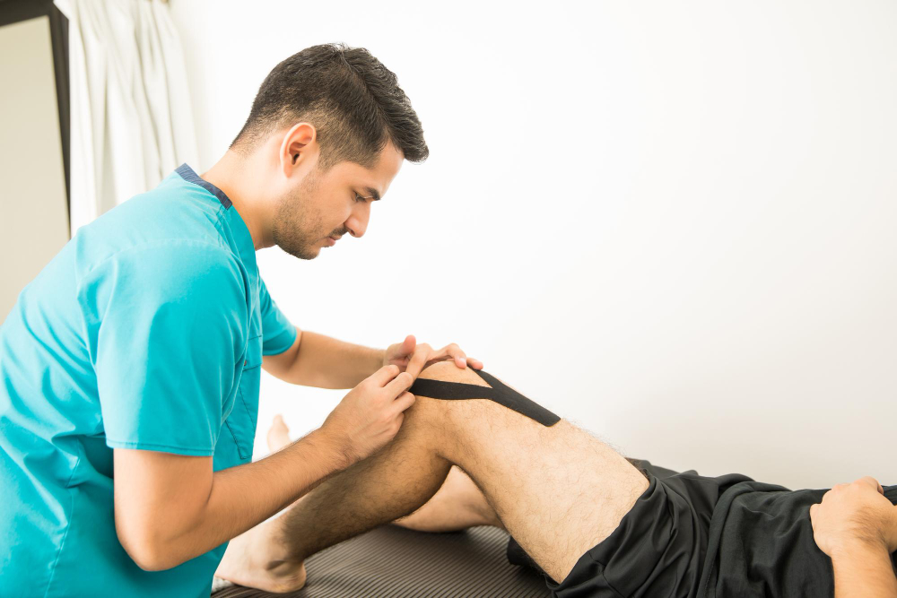
The anterior cruciate ligament (ACL) is a crucial component of the knee that is responsible for offering stability and support throughout the physical exercise. Injury to the ACL occurs frequently, particularly for athletes. They could severely affect flexibility and the quality of your life. When you have the proper ACL injury physiotherapy treatment in gurgaon, the recovery process isn’t just feasible but also efficient and thorough. Understanding ACL Injuries ACL injuries can occur due to ligaments being stretched out or strained due to abrupt changes in direction, abrupt stops or severe impacts. The symptoms typically include swelling, pain, or instability of the knee. The diagnosis of the presence of an ACL injury is usually a matter of physical examinations, imaging such as MRIs and a review of the patient’s flexibility. The Importance of Physiotherapy in ACL Recovery The role of physiotherapy is crucial in the rehabilitation of ACL injuries. It ensures that knees recover strength, stability and flexibility. Highly skilled physiotherapists offer specific exercises and treatments specifically designed to help restore function and stop the recurrence of injury. If you are looking for ACL physiotherapy in Gurgaon, obtaining an organized program that is supervised by experts is vital to ensure successful rehabilitation. Key Components of ACL Injury Physiotherapy Treatment Pain and Swelling Management: It is important to first concentrate to reduce swelling and pain. Methods like compressing, cold therapy as well as elevation, are frequently utilized in conjunction with gentle exercises in order to keep mobility. Strengthening and Stability Exercises: The strengthening of the muscles surrounding the knee, specifically those of the quadriceps as well as the hamstrings is crucial. These exercises assist in strengthening the ligament injured as well as improve the overall stability of your knee. Flexibility and Range of Motion Restoration: The knee’s full mobility is a crucial part of rehabilitation. Joint mobilization and stretching methods ensure that the knee is functioning without issue. Proprioception and Balance Training: ACL injuries are known to impair proprioception, which is the body’s ability to sense joint positions. Training in balance, for example being on one foot or utilizing balance boards can help rebuild this essential component. Return-to-Sport Programs: To help athletes train, certain programmes ensure that the knee is able to handle the requirements of their sports. They include specific exercises for sport, such as agility training and exercises to build strength. Why Choose ACL Injury Physiotherapy Treatment in Gurgaon? Gurgaon is the home of some of the latest rehabilitation centers as well as skilled experts who are experts on ACL rehabilitation. If you’re suffering from the effects of a partial tear or recovering from surgery, our expertise will provide you with individualized care that is tailored to your needs. Clinics that provide ACL physiotherapy services in Gurgaon utilize evidence-based practices and modern technology to aid in quicker healing. Tips for a Successful Recovery Be sure to follow the physiotherapist’s instructions: Consistency and adherence to the plan are essential. Be patient and persistent: ACL recovery takes some time. However, persistence can yield outcomes. Concentrate on Prevention When you are back, keep doing the exercises for flexibility and strength to prevent further injury. Recovery after an ACL injury could be an uphill task, however, when you have the proper assistance and knowledge of ACL rehabilitation physiotherapy in Gurgaon, it is possible to get back to full function and go back to daily routines or sports with a sense of conviction. Do not let an injury stop you back. Research the top treatments for your physiotherapy needs in Gurgaon to ensure successful rehabilitation. What’s the purpose of physiotherapy ACL rehabilitation? The physiotherapy program helps to restore knee strength, stability and mobility. This ensures total rehabilitation. What is the duration of ACL physical therapy typically? Time to recover varies but typically can range between 6 and 12 months in order to fully return to activities that require high impact. Can ACL injuries be repaired with operation? Mild ACL injuries are able to heal by only physiotherapy. However, serious tears typically need surgical treatment and rehabilitation.
Today, in a fast-paced environment, many people are turning to alternative treatments in search of a more healthy and natural method of health and wellbeing. If you’re living in Gurgaon seeking effective alternatives, finding reputable doctors will help in your quest to healthier living. This is a quick guideline to help you find reliable practitioners of alternative medicine in Gurgaon for holistic healing. What Are Alternative Medicines? Alternative medicine encompasses a range of therapies that include Ayurveda, Homeopathy, Acupuncture, Naturopathy, and Yoga. Contrary to conventional medical practices, the treatments are focused on the root causes of illness, not just signs. It doesn’t matter if it’s chronic pain, anxiety, or other lifestyle issues. Alternative therapies offer an avenue to a holistic approach to wellness. Begin by doing some research. The first step towards finding trustworthy alternative medicines in Gurgaon near me, is to conduct some investigation. Utilize online platforms such as Google Reviews, Practo, or Justdial to search for highly rated health centers and doctors. Reviews and reviews can help you understand the kind of experience you can anticipate. Ask for Recommendations One of the most effective ways to identify reputable doctors is to talk with relatives, friends, or even colleagues. If anyone you know is happy when dealing with an individual who offers alternative medicine in Gurgaon there is a good chance you’ll be able to have a positive experience as well. The recommendations of friends and family can save the time and energy spent in the search. Use Online Tools There are numerous applications and websites that help patients connect with health professionals. Applications such as Practo and Zocdoc let you filter your search according to a specific location, the type of treatment as well as reviews from patients. Just enter “alternative medicines in Gurgaon near me,” and you’ll be presented with a list of local options, complete with information. Visit Wellness Centers in Person After you’ve narrowed down some options, go to the facilities to see the surroundings. Cleanliness of the facility? Do the staff members appear courteous and friendly? This is important for the comfort you feel and the level of care you’ll receive. Personal visits also let you talk about your particular concerns directly with the doctor. Check Credentials Be sure to ensure that any practitioners that you choose to consult are licensed and certified. If it’s a holistic practitioner, an ayurvedic physician or a Naturopath, the credentials of the practitioner are important. It is a way to ensure that you’re in good hands as well as receiving professional care from a qualified professional who specializes in alternative medicine located in Gurgaon. Attend Wellness Events Gurgaon is the home of numerous health workshops as well as community gatherings with a focus on the holistic healing process. They are ideal to meet practitioners and learn about different therapies. Some offer complimentary or discounted sessions that allow you to research different treatments with no commitment. Choose What Feels Right It is also essential to locate a center or practitioner which is in alignment with your particular requirements and objectives. Make sure you trust your gut and prioritize your own comfort. Finding the right one can make an enormous difference in the healing process. Locating reliable alternative medicine to use in Gurgaon involves conducting your research by asking friends for suggestions and relying on professionals who have established expertise. With these suggestions, you’ll be in the process of finding the most effective holistic therapies that fit your preferences. What does “alternative” medicine mean? It covers treatments that include Ayurveda, Homeopathy, Acupuncture, Naturopathy, and Yoga which focus on healing that is holistic and natural. How can I determine if the practitioner of alternative therapies in Gurgaon is trustworthy? Make sure they have their certificates. Read online reviews and ask for recommendations from reputable sources. Can alternative drugs available found in Gurgaon in my area be effective? If you have the appropriate practitioner and method, alternative medicine is able to address different health issues effectively.
Stroke is among the most frequent causes of disabilities, and the consequences could be devastating. Regaining mobility is often a matter of the regaining of the ability to move, cognition as well as the capacity to carry out daily tasks. This is the reason why Neuro Physiotherapy Services in Gurgaon can play an important part. Specialized therapies are focused on increasing strength, mobility as well as overall function in patients who have suffered from stroke, giving them an improved level of living. Understanding Neuro Physiotherapy Physiotherapy therapy is a special area of physiotherapy that focuses on helping patients suffering from neurological disorders, including the spinal cord, injuries to the brain, as well as Parkinson’s disease and MS. This therapy is intended to activate nerves, encourage neural development and train the brain to be able to complete actions and perform jobs. Why Choose Neuro Physiotherapy for Stroke Rehabilitation? The patients who suffer from stroke often have difficulties like weakness of muscles as well as impaired balance and diminished coordination. Neuro Physiotherapy Services in Gurgaon near me, have customized treatment programs to address specific problems. The therapists employ evidence-based methods, including: Gait Training to help improve walking. Exercises in balance to avoid falls. Retraining for functional purposes for independence, regaining for daily tasks. They not only improve physical healing, but they also make a positive impact on psychological well-being. Benefits of Neuro Physiotherapy for Stroke Patients Restoring Motor Functions The nervous system’s ability to regulate movement. Neurophysiotherapy aids in the reactivation of the affected muscles and improves the coordination of these muscles through repeated and specific exercises. Reducing Spasticity The muscles stiffness and spasms can be typical post-stroke signs. Physical therapists from Gurgaon employ the stretching technique, massage therapy and electrotherapy to relieve stiffness and increase mobility. Enhancing Balance and Coordination They can cause problems with a person’s balance and coordination, which makes the daily routine difficult. Neurophysiotherapists focus on balance and proprioceptive exercises in order to improve stability and decrease the chance of falling. Why Opt for Neuro Physiotherapy Services in Gurgaon? Gurgaon is the home of one of the most skilled neurophysiotherapists that combine their professional expertise and cutting-edge technologies such as robotic therapy, virtual rehabilitation using augmented reality, as well as hydrotherapy. You may be looking for neuro physiotherapy services in Gurgaon close to my home or are seeking the best medical treatment for a family member. You’ll be able to find a wide range of treatments that cater to your needs for rehabilitation after a stroke. Tailored Programs for Every Patient Every stroke situation is distinct and requires a bespoke method of treatment. Neuro physiotherapy at Gurgaon is a focus on individual assessment as well as goal-oriented therapy programs making sure that each patient’s needs are met. What’s the main focus of physiotherapy for neuro-stroke rehabilitation? The primary focus is the restoration of motor skills, enhancing mobility, and increasing the patients’ independence during everyday routine. What is the length of time that neuro physiotherapy for stroke rehabilitation is? The duration of rehabilitation is contingent upon the degree of the stroke as well as the goals of recovery for the patient; however, a consistent approach generally shows improvements within a few several weeks.
Quality Treatment With Super Affordable Price
Call us anytime