Quality Treatment With Super Affordable Price
Best Physiotherapy Treatment India
Call us anytime
Write a mail

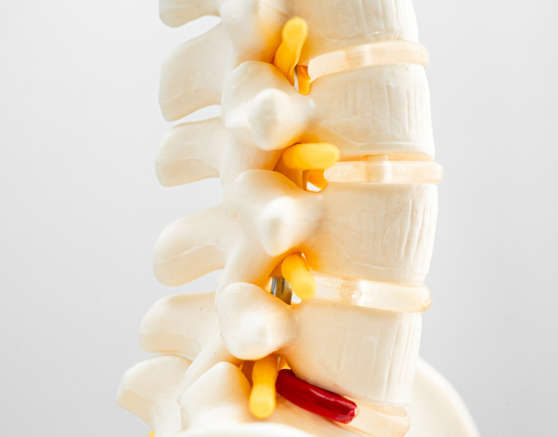
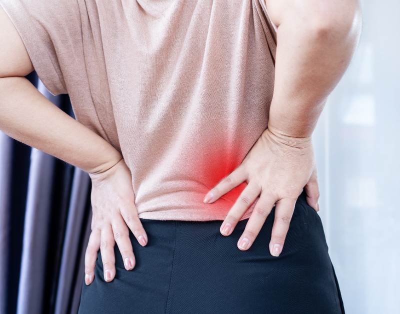
A herniated disc is a frequent spine problem that produces pain and other symptoms. It happens when a gel-like material bulges from a spinal disc, compressing adjacent nerves. This disorder is also known as a slipping disc and, in some instances, a prolapsed disc. Most herniated discs in Gurgaon may be resolved with rest and recovery as the initial therapy. Herniated Disk Or Slipped Disc Causes and risk factors Discs are formed of a robust outer shell with a gel-like center between your spinal column’s bones (vertebrae). The gel in the discs allows the spine to bend and flex while also acting as a shock absorber between the vertebrae. The discs grow less flexible and stiffen as we age, making them more prone to rips. A herniated disc in Gurgaon might develop due to a single severe strain or injury. However, as disc degeneration proceeds with age, some persons may create ruptured discs from modest songs or twists. The following factors can increase the probability of a herniated disc: A herniated disc is most commonly diagnosed between 30 and 50. Men are impacted nearly twice as frequently as women. Herniated Disk Or Slipped Disc Signs and symptoms Some people may have a herniated disc yet have no symptoms. Others suffer from severe, incapacitating symptoms. The location of the herniated disc determines the symptoms encountered. Herniated discs are most commonly found in the lower back (or lumbar spine). Sciatica is the most prevalent symptom of a herniated disc. It is usually an acute shooting pain that runs down the back of one leg from the buttocks. Other symptoms that may occur as a result of a herniated disc include: Bowel or bladder difficulties might indicate cauda equina syndrome, an uncommon but deadly consequence of a herniated disc. If this is suspected, seek immediate medical assistance. Diagnosis Magnetic resonance imaging (MRI) or computed tomography (CT) scanning and X-rays are routinely used to confirm the diagnosis of a herniated disc. A myelogram, which involves injecting dye into the fluid around the spine before taking X-rays, can reveal pressure on the spinal cord or nerves caused by a herniated disc. Nerve conduction examinations, which monitor nerve impulses to the muscles, may also be advised. Non-surgical treatment A herniated disc is often treated with rest, pain medication, and physiotherapy. Bed rest is typically only suggested for up to two days after the beginning of discomfort. More extended rest periods are ineffective at hastening recovery and may even delay it. Keeping up as much mild activity as possible while avoiding unpleasant postures or motions is advisable. A cold compress on the affected area may reduce discomfort and inflammation when symptoms first appear. Applying mild heat to the afflicted area after a few days may relieve pain. Paracetamol and ibuprofen are two medications regularly used to treat the pain associated with a herniated disc. Muscular relaxants may also be administered to treat muscular spasms. Corticosteroid pills or injections may be prescribed in some circumstances. A combination of steroids and anesthesia may be injected directly into the spine for acute pain when other treatments have failed. To preserve mobility and improve the muscles in the back, a mix of physiotherapy treatments and specialized exercises will be performed. Visit Gold Medal Physiotherapy at home treatment for slipped disc. Surgical treatment Surgery may be suggested if non-surgical therapy fails to reduce or eliminate discomfort. If surgery is being considered, it is critical to address the benefits and drawbacks of the procedure, as well as the risks associated with the doctor. Herniated disc surgery may comprise the following methods: All or part of the injured disc is removed to alleviate pressure on the spinal nerves. A discectomy can be “open” surgery (open discectomy) or “minimally invasive surgery” (microdiscectomy). The lamina of the vertebrae is removed to allow more excellent room for the spinal nerves, alleviating pressure on them and lowering discomfort. The disc is removed, and individual vertebrae are fused to minimize mobility. Spinal fusion is a procedure that stabilizes the spine and reduces strain on the spinal nerves. Prevention A herniated disc can be avoided by making the following lifestyle changes: PHYSIOTHERAPY TREATMENT FOR HERNIATED DISK OR SLIPPED DISC Gold Medal Physiotherapy is the Best Physiotherapy Treatment at Home; visit our website and book an appointment.

How To Treat Quadriceps Muscle Strain With Physiotherapy? A quadriceps muscle strain occurs when there is an abrupt tear in the quadriceps muscle in the thigh. It can happen when the power is subjected to severe stress, such as sharp contraction and extension, resulting in the tearing of numerous thigh muscles. Depending on the severity of the muscle rips, numerous treatment options, care, and physiotherapy may be pursued for a quick recovery from quadriceps discomfort. Getting the treatment from the best Physiotherapy for Quadriceps Muscle Strain in Gurgaon is very important. Quadriceps Muscle Strain Different Grades The severity of the damage or tear can be classified into grades. They are as follows: When there is little to no tension, and the quadriceps muscle discomfort is relatively minor, the condition is classified as grade 1. There is usually slight pain, which might make walking and jogging difficult. There is typically no need for rest or further care, and the condition can be treated with brief periods of relaxation between activities. In this grade, no medical intervention is required. When someone engages in an activity such as running or jogging, they experience severe discomfort. It can also result in stiffness and swelling in the quadriceps, making it difficult to bend the knees or cause acute pain. People who cannot move or activate their legs suffer from Grade III agony. The pain in the thighs and leg area is intense, causing tremendous difficulty when walking. There may also be substantial bruising surrounding the area. Quadriceps Muscle Strain Causes Physical activity is one of the most common reasons people get hurt; a sudden impact or the sudden contraction and expansion of muscles can lead to tension and injury. High-performance athletes, such as runners, marathon runners, and other high-contact sports, can contribute. Other causes include lifting significant things, which strains the quadriceps, and external events such as accidents and collisions. These can result in localized damage. In rare situations, an injury might be caused by medical disorders such as gout, obesity, or arthritis. Quadriceps Muscle Strain Symptoms The first sign to watch for is discomfort or strong pull in the thighs and legs. It might indicate a thigh injury that needs to be treated. If the damage is severe, one may have a sharp pain in the thigh and an inability to engage the legs in activities such as running, walking, jogging, and other physical activity. Other symptoms include the inability to extend the knee, leg stiffness, edema, and bruising in the thighs and surrounding areas. A muscle tear is indicated by sharp discomfort while making contact with the thigh, standing up, or putting pressure on the thighs and foot. Quadriceps Muscle Strain Diagnosis A medical professional may inspect the region for swelling and bruises. They may also ask you to perform tasks like extending your legs, bending your knees, and standing on your feet. These acts may reflect the seriousness of the damage. Blood tests and imaging procedures such as X-rays and MRI may be performed to locate the injury and assess the level of damage to the affected muscle and tissues. Doctors may also do Ely’s Test, which analyzes rectus femoris spasticity or tightness. The patient is lying down, and the therapist stands alongside him on the side of the leg that will be evaluated. One hand rests on the lower back, the other on the leg’s heel. Then, bend the knee slightly until the heel contacts the buttocks. For comparison, test both sides. The Test is positive if the heel cannot make contact with the buttocks and the hip of the tested side lifts from the table, and the patient feels discomfort or tingling in the back or legs. Quadriceps Muscle Strain Treatment at Home in Gurgaon One of the most common therapy options is offering anti-inflammatory drugs and pain relievers. It also aids muscle repair by improving blood circulation and alleviating pain. Surgery may be performed in rare cases when there is significant damage to the muscles and tissues. However, this is only done in extreme cases, and rest, meditation, and physiotherapy are commonly used to help restore muscular tightness. The RICE method – Rest, Ice, Compression, and Elevation – can help reduce the pain and severity of an injury by combining muscle cooling and tissue protection. It can also be used to ease pain and tension. Quadriceps Muscle Strain Physiotherapy Treatment The RICE technique is one of the most important techniques, as it may considerably reduce the degree of damage in the area. It also helps with future recovery by decreasing pain and boosting mobility. TENS can assist in reducing inflammation and discomfort while desensitizing nerve fibers, which can help reduce pain and swelling. It can also aid in muscle strengthening. Mobilization can also be accomplished by providing gentle pressure to the affected area to alleviate pain, increase blood circulation, and reduce stress on the nerves and muscles. As the power is activated, it can provide pain relief. Stretching exercises must be done cautiously to avoid stiffening the muscles and allow them to perform effectively. Simple activities such as progressively increasing the degree of straightening the knees and bending the legs can be included. Progressive isotonic quadriceps muscle workouts can help strengthen the muscles in the legs and make them less prone to injury. Simple quadriceps warm-up and cool-down exercises can also help with recovery and enable a smooth transition from injury to performance levels. Quadriceps Muscle Strain Recovery Phase It is critical to avoid putting the muscles under undue strain during the recuperation phase. To reach the best level of healing, one can engage in the exercises indicated above at differing intensities based on consultation with the physical therapist and specialists. The RICE recovery technique is also significant since it causes the muscles to repair faster and ensures they can recover. There may be pain and swelling during this period, but there is a constant decrease in the severity and intensity of the injury. It is critical that

Pes Anserine Bursitis: What Is It And How Can You Treat It? The insertion of three muscles at the proximal end of the Tibia is called Pes Anserine. The Sartorius, Gracilis, and Semitendinosus are among these muscles. Their function in the body facilitates knee flexion; however, they may also play a part in Tibia’s internal rotation and protect the knee from Valgus and rotatory stress. It is considered a crucial portion of the knee for tendon reconstruction surgery or steroid injection therapy of Pes Anserine Bursitis at home. The pes anserine tendons are typically utilized as autographs in knee ligamentous reconstructive procedures. This article investigates the causes and consequences of inflammation of the pes anserine bursa, as well as the treatment regimens that have been proven to be effective. What is Pes Anserine Bursitis? Bursae are sac-like cavitation structures lined by synovial tissues in the body. They must cushion the surrounding structures to avoid frictional injury. Bursitis is a medical word that refers to an inflammatory condition in the Bursa. When these tissues become inflamed, the person may experience discomfort, swelling, and redness. Pes Anserine Bursitis is an inflammation of the Pes Anserine Bursa. Individuals suffering from this illness frequently report discomfort in the medial knee and higher tibial region. It is also known as the pes anserine pain syndrome, characterized by medial knee discomfort. This pain may or may not be connected with Bursa inflammation. What is the Cause of Pes Anserine Bursitis? Direct damage to the Bursa, obesity, misuse of the tissues, tight hamstrings, and mechanical derangement are all major causes of Pes Anserine Bursitis. It is also linked to medial knee osteoarthritis. It is an early and common finding in people with Pen Anserine Bursitis. Individuals who engage in more physical activities and sports regularly may be at a higher risk. Running, basketball, and racquet sports raise an individual’s risk of Pes Anserine Bursitis because they cause overuse and accelerate inflammatory activity. Obesity, as previously stated, is a common risk factor for Pes Anserine Bursitis. Furthermore, research has indicated that obese and overweight people are more likely to develop diabetes mellitus. As a result, because people with diabetes are more likely to develop Pes Anserine Bursitis, fat indirectly plays a causal role. Knee osteoarthritis patients already have elevated inflammatory activity surrounding their knee joints. This inflammation not only promotes the Pes Anserine Bursa but may also result in additional knee issues and structural abnormalities, eventually leading to these Bursa’s inflammation. As previously said, medical derangement is another component that contributes to the occurrence of Pes Anserine Bursitis. It is hypothesized that the medical condition affecting the Medial Knee joint causes increased inflammatory activity in the surrounding hard and soft tissues. Medial meniscus protrusion and medial collateral ligament (MCL) displacement are examples of medical derangements. What are the Symptoms of Pes Anserine Bursitis? Individuals suffering from Pes Anserine Bursitis in Gurgaon frequently complain of discomfort inside the knee, also known as the medial aspect. This discomfort is generally provoked by sitting, stepping on stairs, or crossing one’s legs. When people cross their legs, three muscles known as the semitendinosus, Gracilis, and Sartorius act together, which is why this posture frequently causes discomfort, as observed in Pes Anserine Bursitis. Some people may also have muscular weakness or a decrease in the range of motion of the knee joint. Others have reported soreness while pressing the skin over the insertion of the Pes Anserine Tendons. It is also known as the goose’s foot and is found between the upper medial Tibia and the medial knee. Some people may suffer edema as a result of inflammatory activity. The doctor may ask the patient to perform knee flexion at a 90-degree angle to check the soreness’s presence. The individual may feel discomfort when the medial tendinous structure along the proximal medial tibial area is palpated in this posture. How is Pes Anserine Bursitis Diagnosed? Bursitis of the Pes Anserine Clinical examination is used to make the diagnosis. While lab testing is typically not required, a health practitioner may use imaging technologies to provide a more accurate diagnosis. These imaging techniques should always be used with a comprehensive medical history and physical examination. When a patient complains of severe pain, the doctor may recommend an x-ray to confirm no fractures or foreign body incursions. When a healthcare practitioner has to distinguish between inflamed Bursa and cellulitis, ultrasonography may be a superior imaging technique. It may allow for a live investigation of the range of motion and eliminate any tendinous injuries. Some people may be advised to get Magnetic Resonance Imaging. However, this is uncommon. It might rule out prepatellar Bursitis, an oval fluid-filled lesion between the knee joint and soft tissues. The Recommended Treatment Options for Pre-Anserine Bursitis To establish the most appropriate treatment strategy for the patient, the causes of Pre Anserine Bursitis should be thoroughly explored. Systemic medication therapy should be used initially if the reason is a medical illness such as Gout. Antibiotic treatment may be recommended if the cause of infection is identified in septic Bursitis. It is followed by a general care strategy for all varieties of Bursitis, which includes resting, icing, modifying activities, and taking anti-inflammatory drugs. Overall, therapeutic approaches may be classified into conservative care and surgical management. While most acute instances of Pre Anserine Bursitis may be managed conservatively, certain severe cases may necessitate surgical intervention. Chronic instances of Pre Anserine Bursitis may require corticosteroid injections rather than over-the-counter treatment. These patients are also advised to return to the doctors regularly to track their development and determine which therapies may be most suited for them at this time. Medical Treatment The first step in beginning medical therapy for pes anserine Bursitis is the prescription of an over-the-counter Nonsteroidal anti-inflammatory medicine (NSAID). However, if it is not proven to be useful for pain alleviation and unpleasant symptoms of the individual, alternative techniques such as steroid injections may be shown to be effective. The second-line therapy


Quality Treatment With Super Affordable Price
Call us anytime
